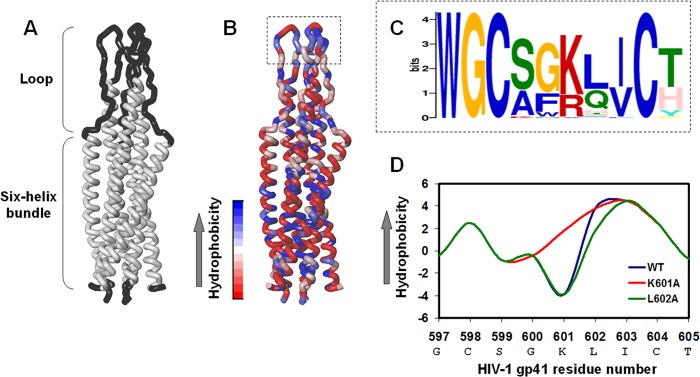
FIGURE 1.

Bioinformatics characterization and mutagenesis of the hydrophobic loop core. A, presentation of the loop structure devoid of the cysteine residues together with the six-helix bundle in the soluble hairpin conformation of the SIV fusion protein. This is the only available structure of the loop. The three-dimensional structure was taken from Caffrey et al. (13), PDB ID 1QCE. B, the location of the hydrophobic core within the loop region. The highest hydrophobic residues are blue, and the lowest hydrophobic residues are red. C, sequence homology in the loop hydrophobic core between HIV and SIV clades shows conservation of the basic residue (Lys or Arg) between the two Cys residues. D, alteration of the hydrophobic level of the gp41 loop core by mutagenesis analysis. Residue numbers correspond to the gp160 HIV-1 HXB2 variant.
