
FIGURE 2.
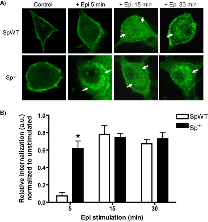
Epinephrine-mediated α2AAR endocytosis is regulated by spinophilin. A, neurons with (SpWT) and without (Sp−/−) spinophilin expression were stimulated by application of Epi (100 μm, plus prazosin and propranolol) as indicated. Neurons were subjected to α2AAR prelabeling method as in Fig. 1 (B and C). Endocytosis, indicated by the appearance of intracellular punctae containing internalized receptors (arrows), was observed beginning at 5 min in Sp−/− neurons and at 15 and 30 min in both SpWT and Sp−/− neurons, consistent with endocytic acceleration in the absence of spinophilin. B, quantitation of agonist-mediated α2AAR endocytosis in Epi-stimulated SpWT and Sp−/− neurons, with relative internalization determined as described under “Experimental Procedures.” Confocal images are representative of at least three independent samples, and quantitation was performed over at least 12–14 neurons. *, p < 0.01, SpWT versus Sp−/−.