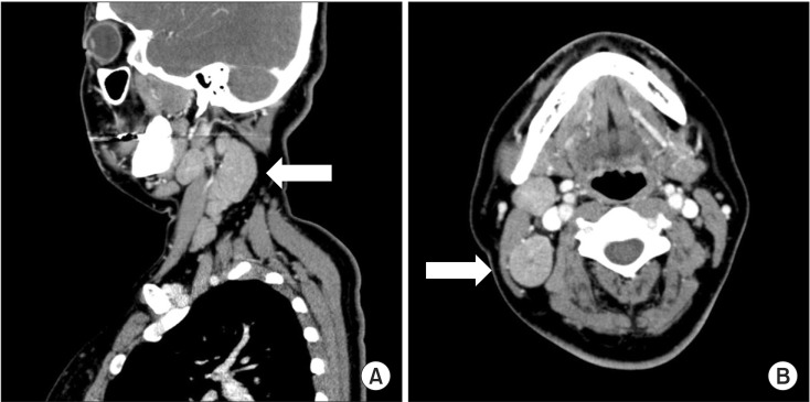
Figure 1.
Sagittal (A) and axial (B) images of enhanced neck computed tomography (CT). (A) Sagittal images of enhanced neck CT show multiple enlarged lymph nodes at levels II, III, IV, and V. (B) Axial images of enhanced neck CT show multiple enlarged lymph nodes at levels II, III, IV, and V. The short diameter of the largest lymph node is approximately 3 cm (arrow).

