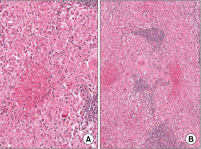
Figure 2.

Microscopic findings of the case. (A) An epithelioid granulomatous lesion with central necrosis and multinucleated giant cells is present (H&E stain, ×100). (B) High magnification of (A) shows hyalinized necrosis and surrounding epithelioid cells (H&E stain, ×200).
