
Figure 1.
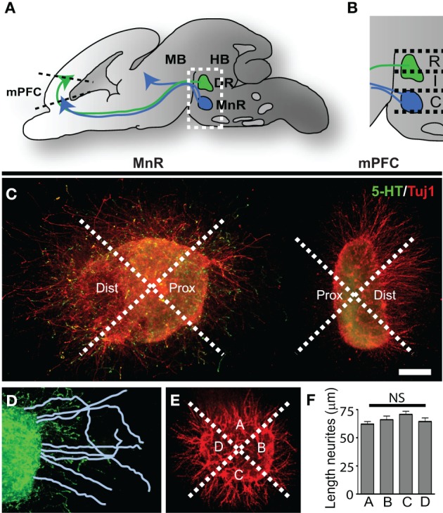
Three-dimensional collagen co-cultures of explants taken from the mPFC, DR, and MnR show trophic responses. (A) Schematic of an embryonic brain showing the position of the 5-HT-positive rostral cluster of raphe nuclei projecting to the mid- and forebrain. The dorsal raphe nucleus (DR) projects to forebrain regions (green arrow) including the prefrontal cortex (mPFC). The median raphe nucleus (MnR) projects (blue arrows) to fore-and midbrain regions. (B) Enlargement of the boxed area in (A). The rostral (R) and intermediate (I) subarea correspond to the DR and the caudal (C) subarea corresponds to the MnR. (C) Example of a 5-HTT+/− caudal subarea (MnR) co-cultured with mPFC, divided in proximal and distal quadrants and stained for 5-HT (5-HT neurites, green) and Tuj1 (β-III tubulin, all outgrowing neurites, red). (D) In the proximal (and distal, not shown) quadrants the neurites are traced and measured. (E) Control explants were cultured separately and neurite outgrowth was measured in the 4 quadrants (example of WT mPFC). (F) The average length of the neurites in quadrants A, B, C, or D showed no significant (NS) difference. HB, hindbrain; MB, midbrain. Scale bar represents 80 μm.