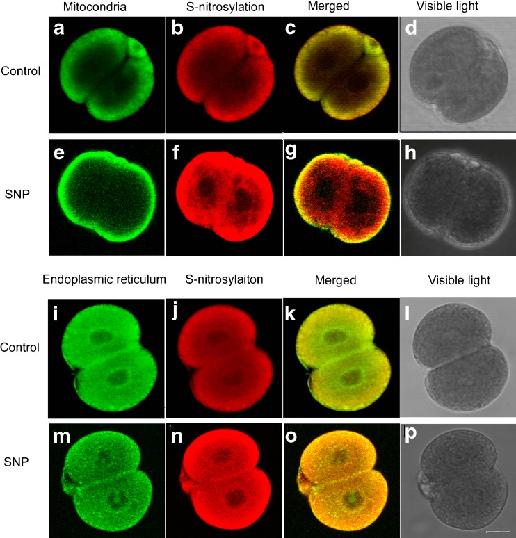
Fig. 3.
Patterns of protein S-nitrosylation in 2-cell mouse embryos following NO donor (10 μM SNP) exposure were observed using confocal microscopy. a and e Mitochondria were stained with MitoTracker Green FM and the mitochondria of 2-cell mouse embryos were mostly located in the cortical area of the cytoplasm both in control and NO exposure group; i and m The endoplasmic reticulum (ER) was stained with Con-A; b, f, j and n S-nitrosylated proteins were stained using the biotin switch method and MTSEA-Texas red. f vs. b and n vs. j Protein S-nitrosylation increased post-NO exposure, and extended from the cortical area toward the nucleus. c and g S-nitrosylated proteins were more colocalized with mitochondria in the control group compared to SNP-treated group. k and o S-nitrosylated proteins closely colocalized with ER, prominently post-NO exposure. The bar in p indicates 20 μm. Images were captured using a Leica TCS NT microscope with a 40× objective and NA 0.55

