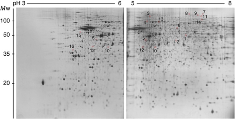
Figure 1.
2-D proteome reference maps obtained using narrow pH range IPG strips. Proteins of mouse hypothalamus (200 μg) were separated on pH 3–6 and pH 5–8 IPG strips in the first dimension and by 13% SDS–PAGE gels in the second dimension. Proteins were stained with SYPRO Ruby. Differentially expressed spots (P<0.02) that are labelled and numbered were identified by LC-MS/MS (see Table 2 and Supplementary Tables 1 and 2). The spots 5, 10, and 12 were found independently in gels with both kinds of pH range.

