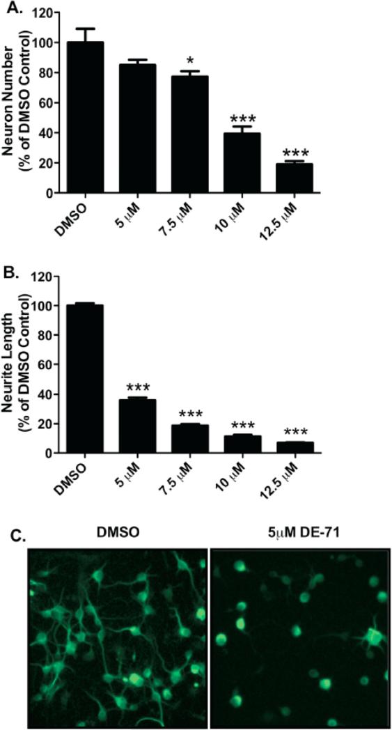
Figure 2.
Exposure of cortical primary cultures to DE-71 causes a loss of cortical neurons as well as reductions in neurite outgrowth. (A) Treatment of cortical cultures caused a significant reduction in the number of MAP2+ neurons, compared with DMSO control (52.3 ± 11.7) at 7.5 (40.5 ± 4.6), 10 (20.6 ± 6.0), and 12.5 μM (9.9 ± 2.8) DE-71. (B) Assessment of neurite length in these neurons demonstrated a greater loss of neurite outgrowth with a substantial reduction, compared with DMSO control (45.3 μm ± 1.8) at 5 μM (16.3 μm ± 2.0), 7.5 μM (8.5 μm ± 1.0), 10 μM (5.1 μm ± 1.4), and 12.5 μM (3.2 μm ± 0.4) DE-71. Columns represent the percent change from DMSO control for each genotype. Data represent the mean ± SEM of 4 experimental replicates per treatment group performed across 3 separate experiments. *Values for treatments significantly different from their respective genotype DMSO control (p < 0.05). ***Values significantly different from their respective genotype DMSO control (p < 0.001). (C) Representative cortical cultures stained for MAP2 and treated with DMSO or 5 μM DE-71.

