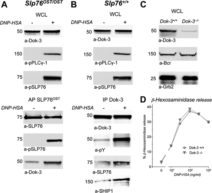
Fig. 6.
Validation of the SLP76-Dok-3 interaction in activated mast cells. A, Co-purification of Bcr with SLP76OST. Slp76OST/OST BMMC sensitized with IgE anti-DNP were challenged with DNP-HSA (+) or without (−) for 2 min at 37 °C and lysed. Samples of whole cell lysates (WCL) were Western blotted with anti-Dok-3 antibodies (upper panel). Remaining lysates were subjected to affinity-purification with Strep-Tactin. Eluates were electrophoresed and Western blotted with anti-SLP76, anti-phosphotyrosine (pY), and anti-Dok-3 antibodies (lower panel). B, Co-immunoprecipitation of SLP76 and SHIP1 with phosphorylated Dok-3. Slp76+/+ BMMC sensitized with IgE anti-DNP were challenged with DNP-HSA (+) or without (−) for 2 min at 37 °C and lysed. Samples of whole cell lysates (WCL) were Western blotted with anti-Dok-3 or anti-phospho-PLCγ-1 antibodies (upper panel). Remaining lysates were immunoprecipitated with anti-Dok-3 antibody. Eluates were electrophoresed and Western blotted with anti-Dok-3, anti-phosphotyrosine (pY), anti-SLP76 or anti-SHIP1 antibodies (lower panel). (C, D) Lack of genetic evidence that Dok-3 is involved in FcεRI signaling. Aliquots of BMMC from Dok-3−/− and from Dok-3+/+ mice were lysed in SDS. whole cell lysates (WCL) were Western blotted with anti-Dok-3 antibodies and, as positive controls, with anti-Bcr or anti-Grb2 antibodies (C). Aliquots of the same cells were sensitized with IgE anti-DNP, and challenged with the indicated concentrations of DNP-HSA. β-hexosaminidase was measured in supernatant 10 min later (D).

