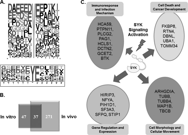
Fig. 4.
A, Consensus sequence analysis of in vitro kinase phosphorylation for SYK specificity. Upper panel, frequency plot of all amino acids flanking the phosphotyrosine site. Lower panel, significantly enriched motif from SYK substrates using Motif-X; B, Venn diagram illustrating the overlap of tyrosine phosphorylation sites identified by in vitro kinase reaction and in vivo phosphoproteomics in DG75 cells. C, Categories of biological processes for identified SYK substrates.

