
Fig. 10.
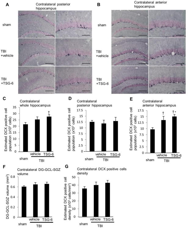
Administration of TSG-6 protein increased neurogenesis in the hippocampus at 10 weeks after TBI. TSG-6 protein (50 μg/mouse) was administered twice at 6 and 24 hour after TBI. (A, B) Images of representative sections showing distribution of newly born neurons expressing doublecortin (DCX) in the subgranular zone-granule cell layer (SGZ–GCL) of contralateral posterior (A) and anterior (B) hippocampus at 10 weeks after TBI from sham and injured mice that received vehicle or TSG-6. Left: ×10 magnification (scale bars=200 μm), Right: ×20 magnification (scale bars = 100 μm). (C-E) Numbers of DCX positive newly born neurons. The total numbers of DCX positive neurons were estimated from unbiased stereological cell counts of the subgranular zone-granule cell layer (SGZ-GCL). Seven sections were collected from the whole hippocampus, one at each 450 μm. The number of DCX positive neurons was calculated from all seven sections (C), posterior three sections (D) or anterior four sections (E). The volume of dentate gyrus (DG) SGZ-GCL was calculated by Stereo Investigator system from seven sections (F). The density of DCX positive cells was calculated as dividing the number of DCX-positive cells by the volume of DG-SGZ-GCL (G). n = 9 or 10 mice/group. All data are represented as mean ± SEM. †p<0.05 versus the sham group, *p < 0.05 versus the vehicle group.