Fig. 5.

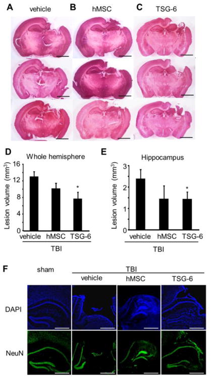
Intravenous injection of TSG-6 protein protected against TBI induced tissue loss in vivo after TBI. hMSCs (106 cells/mouse) were administrated 6 hour after TBI. TSG-6 protein at dose of 50 μg/mouse was administrated twice at 6 and 24 hour after TBI. (A-C) Representative H&E stained sections from injured mice treated with vehicle (A), MSC (B) or TSG-6 (C) at 14 days after TBI. Scale bars = 2 mm. (D, E) Lesion volume of whole hemisphere (D) and hippocampus (E) treated with vehicle (n=8), hMSCs (n=5), or TSG-6 (n=5) at 14 days after TBI. (F) Representative images of NeuN and DAPI stained hippocampus of mice 14 days after TBI. Scale bars = 500 μm. All data are represented as mean ± SEM. *p < 0.05 versus the vehicle group.
