Figure 1.
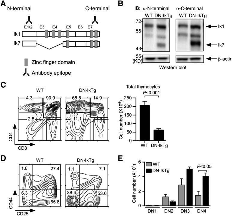
Dominant-negative Ikaros impairs T-cell development in the thymus. (A) Exon organization of full-length Ikaros-1 (Ik1) and the DN splice variant Ik7. E1-E7 corresponds to exon 1-exon 7. Antibody binding sites indicate epitopes of immunoblot antibodies. (B) Immunoblot analysis of WT and DN-IkTg thymocytes. Whole-cell lysates were probed with Ikaros N-terminal (left) or C-terminal (right)–specific antibodies. The same blot was reprobed with anti–β-actin antibodies for loading control. The blots are representative of 3 independent experiments with each 1 WT and 1 DN-IkTg mice. (C) Thymocyte profiles and cell numbers of WT and DN-IkTg mice. Contour plots show CD4/CD8 profiles of total thymocytes (left). The bar graphs indicate the mean ± SEM of total thymocyte numbers (right). Data represent a summary of 11 WT and 13 DN-IkTg mice from 11 independent experiments. (D) CD44 vs CD25 expression in lineage marker–negative, immature DN thymocytes. Contour plots are representative of 4 independent experiments with each 1 WT and 1 DN-IkTg mouse. (E) Cell numbers of individual DN subpopulations. The bar graphs indicate the mean ± SEM of 3 independent experiments with each 1 WT and 1 DN-IkTg mouse.

