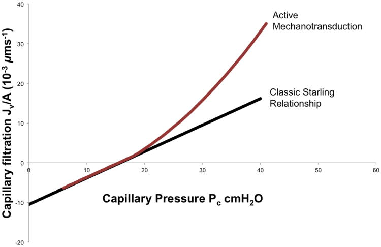
Figure 3.

Schematic illustrating the hypothesized role of the glycocalyx in lung vascular mechanotransduction. Left: during static conditions, the glycocalyx maintains barrier function over the intercellular junction. Right: during increased vascular pressure, the increased hydraulic flow through the glycocalyx deforms or stresses the glycosaminoglycan (GAG) fibers, which in turn activates endothelial nitric oxide synthase (eNOS) and leads to barrier dysfunction. ΔPc, change in capillary pressure; Q, flow; ZO-1 and ZO-2, zonula occludens-1 and -2; vin, vinculin; VE-Cad, vascular endothelial cadherin; ECM, extracellular matrix (Adapted from Dull R, Cluff M, Kingston J, Hill D, Chen H, Hoehne S, Malleske D, Kaur R. Lung heparan sulfates modulate Kfc during increased vascular pressure: evidence for glycocalyx-mediated mechanotransduction. Am J Physiol Lung Cell Mol Physiol 2012;302:L816-28).
