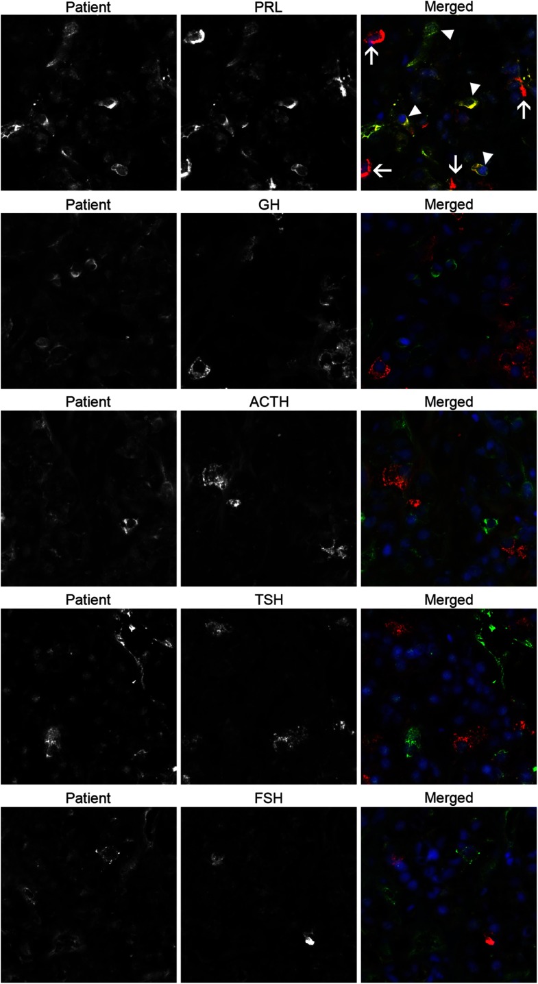
Figure 2.
The patient autoantibodies recognize a specific subset of PRL-secreting cells in double immunofluorescence experiments using human pituitary as the substrate. Each horizontal series of 3 panels represents the staining obtained using only the patient serum (left panels), only a commercial anti-hormone antibody (center panels), or the two combined (right panels). The top 3 panels represent the experiment with the anti-PRL antibody. It shows that the patient antibodies recognize some PRL-secreting cells that are also recognized by the commercial anti-PRL antibody, and thus appear yellows when the two images are merged (top right panel, arrow heads). It also shows that there are other PRL-secreting cells that are not recognized by the patient serum (top right panel, arrows). Performing the same experiments with commercial antibodies to GH (second row), ACTH (third row), TSH (fourth row), or FSH (fifth row) showed no colocalization of the staining.

