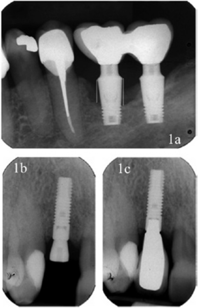
Figure 1.

a) X-ray containing the references used in the measurements made from the implant shoulder to bone level, both mesially and distally. b) X-ray at the time of implant loading, showing that the bone margin coincides with the implant shoulder. c) X-ray after one year of functional loading in a patient who has received supportive periodontal therapy. The maintenance of the bone level can be observed.
