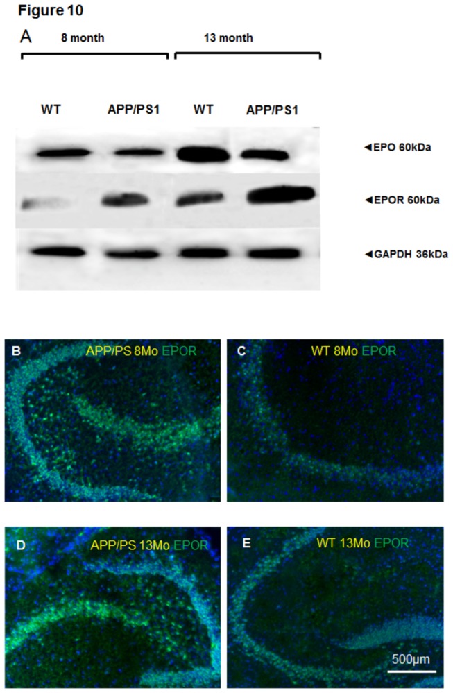
Figure 10. EPO and EPOR expression in 8- and 13 month old APP/PS1 and WT mice.

(A) Western blot analysis of EPO and EPOR in brain homogenates from 8- and 13-month-old APP/PS1 and WT mice. GAPDH serves as a loading control (B) Representative immunostaining of EPOR in the hippocampal area of 8-month old APP/PS1 mouse; (C) EPOR expression in 8 month old WT mouse hippocampus; (D) EPOR (green) in 13 month old APP/PS1 mouse hippocampus; (E) Corresponding WT-control to D showing EPOR (green) in 13 month old WT mouse hippocampus. Scale bar 500µm.
