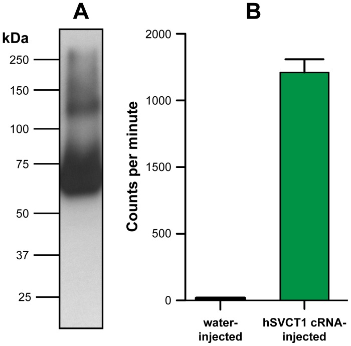
Figure 1. Expression and function of hSVCT1 in Xenopus oocytes.
(A) Western blot analysis of isolated membranes from three oocytes expressing hSVCT1. Western blotting was performed using a 6% SDS/polyacrylamide gel and an anti-HA antibody. The prominent band below the 75 kDa marker corresponds to recombinant hSVCT1 (calculated molecular mass based on the amino acid sequence: ∼ 71 kDa). A second, less prominent band between the 100 kDa and 150 kDa markers was assigned to dimeric hSVCT1. (B) Oocytes injected with hSVCT1 cRNA mediate uptake of [14C]ascorbate in contrast to control oocytes injected with water. Mean ± SEM from 20 oocytes are indicated (two independent experiments).

