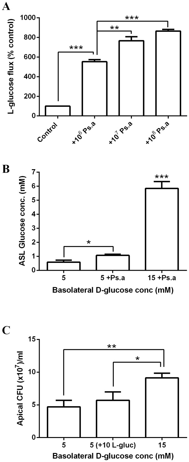
Figure 4. Apical Ps. aeruginosa addition increases glucose flux across Calu-3 airway epithelial monolayers, resulting in elevated ASL glucose and D-glucose dependent bacterial growth.
A: Basolateral-to-apical paracellular L-glucose (14C-L-glucose) flux across untreated Calu-3 monolayers and monolayers pre-exposed to Ps. aeruginosa (1×10⁶, 1×10⁷ or 1×10⁸ CFU) for 7 hours. n = 3 in each group, obtained from 3 independent experiments. ** p<0.01, *** p<0.001. B: Glucose concentration of airway surface liquid (ASL) across Calu-3 airway epithelial monolayers after 24 hours, in the presence of 5 or 15 mM basolateral glucose, with or without Ps. aeruginosa filtrate (Ps. a). n = 3 in each group, obtained from 3 independent experiments. * p<0.05, *** p<0.001. C: Ps. aeruginosa growth over 7 hours across the apical surface of Calu-3 monolayers, in the presence of 5 mM D-glucose, 15 mM basolateral D-glucose or 5 mM D-glucose plus 10 mM L-glucose. n = 6 in each group, obtained from 3 independent experiments. * p<0.05, ** p<0.01.

