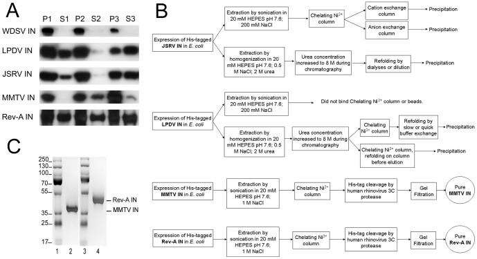
Figure 1. IN expression, extraction, and purification.
(A) Fractions of bacterially expressed His6-tagged WDSV, LPDV, JSRV, MMTV, and Rev-A INs were visualized through western blotting. Lanes 1 and 2 represent the pellet (P1) and supernatant (S1) fractions obtained following centrifugation of cells lysed in 200 mM NaCl-containing buffer A. Pellet 2 (P2) and supernatant 2 (S2) were obtained following centrifugation (lanes 3 and 4) of fraction P1 homogenized in buffer B containing 1 M NaCl and 5 mM CHAPS. During the final extraction step, the pellet from step 2 was homogenized in buffer C containing 0.5 M NaCl and 2 M urea (lanes 5 and 6). (B) Schematic of the protocols utilized for JSRV, LPDV, MMTV, and Rev-A IN purification. All columns were run on an ÄKTA purifier system. (C) The purities of MMTV (lane 2) and Rev-A (lane 4) INs were assessed at 93% and 97%, respectively, following silver staining of SDS-polyacrylamide gels. Lanes 1 and 3 contain the indicated molecular mass standards.

