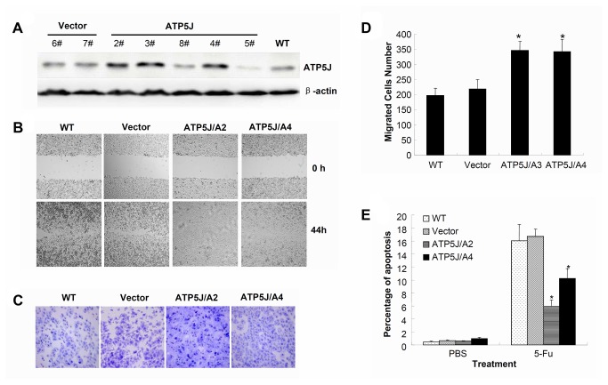
Figure 5. Up-regulation of ATP5J expression enhanced cell migration and decreased 5-Fu sensitivity in DLD1 cells.
(A) Western blot result for ATP5J protein in DLD1 cells after stable transfection with pcDNA3.1(+)/ATP5J plasmid. (B) Wound-healing assay for WT, Vector, ATP5J/A2, and ATP5J/A4 cells. Data represent one of three similar experiments (original magnification: x100). (C) Migrations of WT, Vector, ATP5J/A2, and ATP5J/A4 cells were assayed using a 24-Transwell system. The pictures of migrated cells were taken 24 hours after seeding (original magnification: x100). Data represent one of three similar experiments. (D) Quantitative analysis of three cell-migration assays. Values represent means ± SD. *P<0.05. (E) Apoptotic ratios of WT, Vector, ATP5J/A2, and ATP5J/A4 cells after treatment with 50 µmol/L 5-Fu for 3 days. Data represent one of two similar experiments. Values represent means ± SD and *P<0.05.

