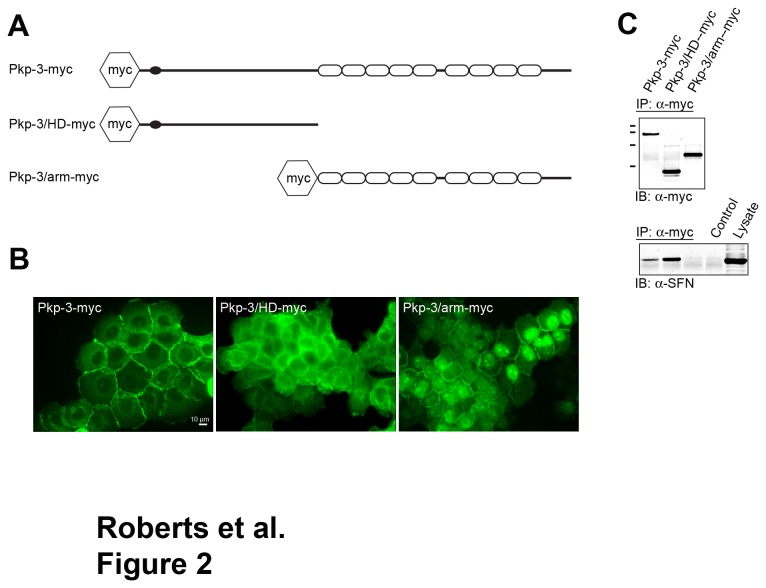
Figure 2. Stratifin interacts with the amino terminal domain of plakophilin-3.
A. Retroviral expression vectors were generated encoding epitope-tagged plakophilin-3 (Pkp-3-myc), plakophilin-3 amino acids 1-307 (Pkp-3/HD-myc) and plakophilin-3 amino acids 308-797 (Pkp-3/arm-myc). B. Immunofluorescence microscopy using an anti c-myc antibody (9E10) revealed the subcellular localization of the exogenous plakophilin-3 proteins stably expressed in A431 cells. C. (upper panel) Immunoblot analysis of plakophilin-3 immunoprecipitated from cell lysates prepared from A431 cells expressing the plakophilin-3 demonstrate the exogenous proteins are immunoprecipitated at approximately equal levels, and the proteins migrate at the appropriate molecular mass by SDS-PAGE (MW markers from top to bottom; 116 kDa, 97 kDa, 68 kDa, and 45 kDa). (lower panel) The immunoprecipitates were immunoblotted using anti stratifin. Note that full length Pkp-3-myc and the Pkp-3/HD-myc co-immunoprecipitated stratifin while Pkp-3/arm-myc did not.

