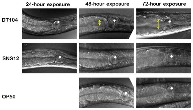
Figure 3. Colonization of the C. elegans intestine by Salmonella Typhimurium DT104.

Confocal microscopy images of representative worms exposed to DT104, SNS12, and E. coli OP 50. White arrow shows the grinder of the pharynx, and yellow line shows extent of intestinal distention.
