
Figure 1. a) Representation of domain structure of sub-classes of MetAP based on the crystal structures.
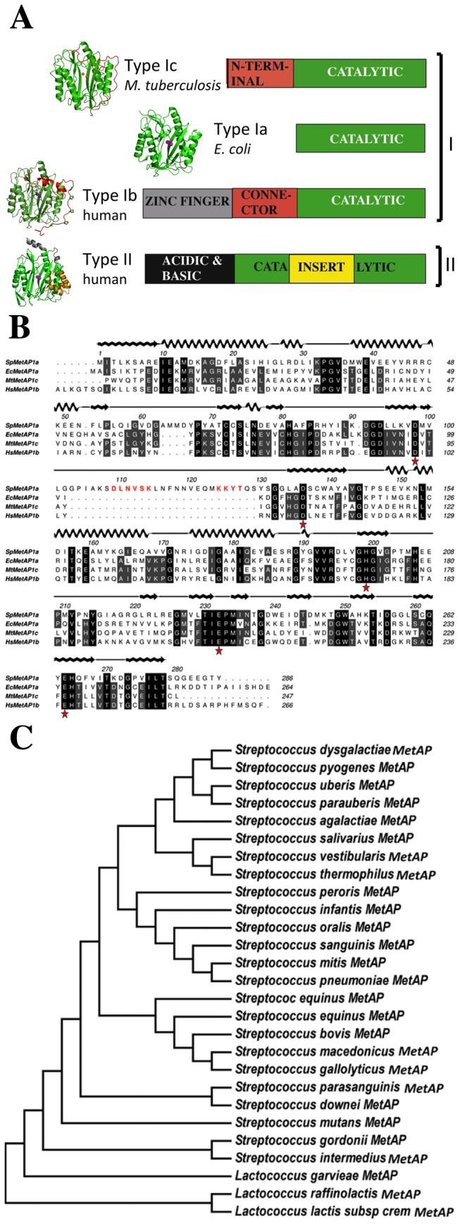
Common catalytic regions are shown in green. Type Ia has the basic structure required for catalysis. Type Ib and Type Ic have extra regions on the amino terminus. Type II MetAP has a 60 amino acid insert in addition to N-terminal extension (yellow). On the left of each bar is the representative crystal structure in cartoon diagram. Pink spheres in the middle indicate the two metal ions in the active site. b) Sequence alignment of Type I MetAPs from S. pneumoniae (Type Ia), E. coli (Type Ia), M. tuberculosis (Type Ic) and human (Type Ib). Numbering and the secondary structure representation on the top of the alignment are based on the SpMetAP1a crystal structure. Metal binding conserved residues are marked as asterisk. Two inserts are identified63; insert and 103insert. These inserts are predominantly present in the streptococcus bacterial Type Ia MetAP109. DLNVSK and 124KKYT are two sequences marked in red are predicted to have respectively glycosylation and phosphorylation sites. c) Phylogenetic tree of 24 Type I MetAPs from streptococcal and lactococcal bacteria that contain the two inserts observed in the Figure 1b. Note that each of these species forms separate clusters.