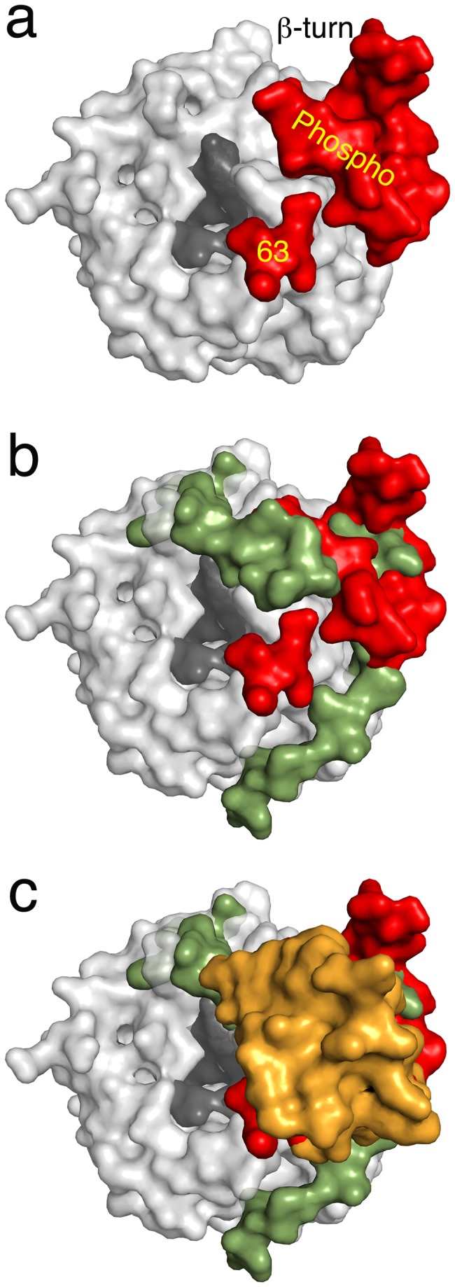
Figure 4. Genetic variations of MetAP’s with additional sequences.

Catalytic domains are shown in light grey color while the entrance to the active site in the dark grey. a) Crystal structure of SpMetAP1a. Two extra inserts are depicted in red. b) Overlay of SpMetAP1a and MtMetAP1c. Extension of the N-terminus of the MtMetAP1c is shown in green. The inserts in the SpMetAP1a and N-terminal extension in the MtMetAP1c structurally align well suggesting of common function. c) Overlay of SpMetAP1a, MtMetAP1c and HsMetAP2 structures. The insert domain of the later enzyme is shown in gold color. Note that all three extra modifications on the three different MetAP’s align at the same place reconfirming the common functional roles of these extra regions.
