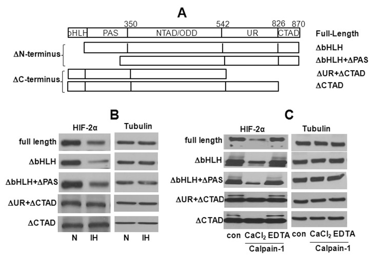
Figure 7. Involvement of N- terminus and C- terminus HIF-2α in IH-induced HIF-2α degradation by calpains.
A. Schematic diagram showing the full length, N-terminus (ΔbHLH and ΔbHLH+ΔPAS) and C-terminus (ΔUR, ΔUR +ΔCTAD) deleted constructs. B. Western blot showing HIF-2α protein in PC12 cells transiently transfected with the HIF-2α full length and the two N- and C-terminus truncated constructs and exposed to normoxia (N) or IH. The N-terminus and C-terminus deleted HIF-2α proteins were detected with antibody raised against HIF-2α N-terminus (Acris Antibodies; AP23352PU-N) and C-terminus (Novus Biologicals; NB100-122) respectively. C. PC12 cell lysates expressing the N- and C-terminus deleted protein were incubated for 15 min with purified calpain-1 (3 mg/ml) in presence of 1 mM CaCl2 or 1 mM CaCl2+2 mM EDTA and HIF-2α protein was analyzed by western blot.

