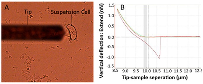
Figure 1. Stiffness-properties imaging by atomic force microscopy (AFM) technology.

(A) Optical image of a typical single rice cell sample. The shadow of the AFM cantilever is visible on the left-hand side of the image. (B) A typical force-distance (FD) curve recorded on a single rice cell sample. The upper line represents the FD curve when the tip indents (or penetrates) the cell wall and the lower line represents the FD curve when the tip retracts from the cell wall.
