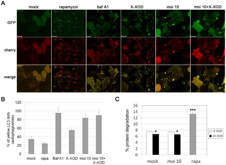
Figure 4. Oxidative stress and HSV-1 infection induced inefficient fusion between autophagosomes and lysosomes.
A) SK-N-MC cells were transfected with dtLC3 and then infected with HSV-1 at a moi of 10 for 18 h, or treated with bafilomycin A1 (baf A1), rapamycin (rapa) or X-XOD for 18 h. Representative confocal microscopy images are shown; the GFP (green) and mCherry (red) channels were merged. Scale bars: 10 µm. B) Graphic representation of the proportion of autophagosomes (yellow dots). The number of fluorescent bodies in 100 cells was counted for each condition. C) Degradation of long-lived proteins in SK-N-MC cells infected with HSV-1 at a moi of 10 for 18 h in the presence or absence of X-XOD. Rapamycin (rapa) was used as a positive control of autophagy stimulation. Results are the mean ± SEM of three independent experiments performed in triplicate (*p<0.05; ***p<0.001).

