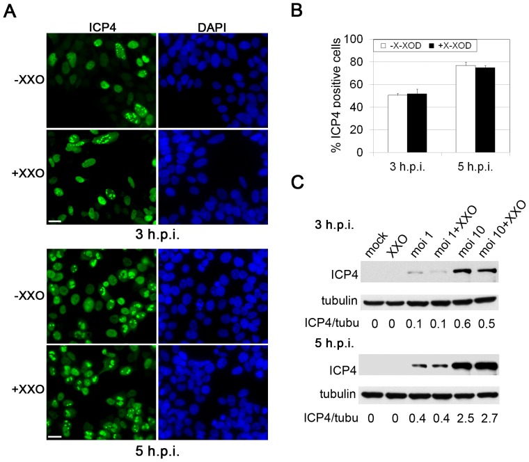
Figure 5. Effects of oxidative stress on HSV-1 entry.
A) Immunofluorescence analysis of HSV-1-infected SK-N-MC cells at a moi of 10 in the presence and absence of X-XOD. The immunoreactivity of ICP4 protein is shown at 3 and 5 hours post-infection (h.p.i.). Nuclei are stained with DAPI. Scale bar: 20 µm. B) Quantification of infected cells by ICP4 staining. The graph shows the percentage of ICP4-positive cells. At least 400 nuclei were counted for each condition. C) Analysis of ICP4 levels by Western blotting in SK-N-MC cells infected with HSV-1 at a moi of 1 and 10 for 3 h and 5 h, in the presence and absence of X-XOD.

