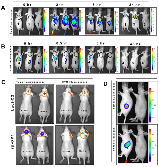
Figure 6. Targeting of subcutaneous tumor xenografts by fluorescently labeled SapC-DOPS nanovesicles.
Heterotopic MiaPaCa-2 tumors (circled) were generated by subcutaneous injection in the upper flank of nude mice. (A) Mice 1 & 2 were tumor-bearing mice, injected with fluorescently labeled SapC-DOPS nanovesicles; Mouse 3 was non-tumor-bearing, PBS injected. Mice 1 and 2 both demonstrate localization of the fluorescent label to the tumor site. Fluorescence also localizes to the liver by 2 hours, but is gone by 24 hours. (B) Tumor-bearing mice 4, 5, and 6 were injected with non-complexed SapC and fluorescently labeled DOPS, fluorescently labeled DOPS only, and PBS, respectively. There is no fluorescence localized to tumor in any of these mice. Like the SapC-DOPS (in mice 1 and 2), non-complexed SapC and fluorescently labeled DOPS and fluorescently labeled DOPS alone (in mice 4 and 5, respectively) did localize to the liver, but quickly dissipated (C) Subcutaneous tumors created using cfPac1-Luc3 pancreatic tumor cells that were and were not pretreated with PS-specific binding proteins (Lactadherin-C2 [left upper panel] and Beta-GP-1 [left lower panel]) display bioluminescence on live imaging. After administration of CVM fluorescently labeled SapC-DOPS nanovesicles, the tumors that were not pretreated demonstrated fluorescence, while the tumors that had been pretreated did not demonstrate any fluorescence (right upper and lower panels). (D) Presence of bioluminescence (upper panel) confirms presence of orthotopic pancreatic tumor, and co-localized fluorescence (bottom panel) confirms targeting by fluorescently labeled SapC-DOPS nanovesicles.

