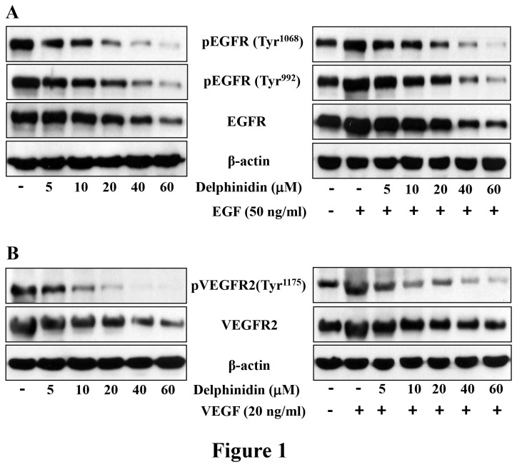
Figure 1. Effect of delphinidin treatment on the constitutive and EGF- and VEGF-induced phosphorylation of EGFR and VEGFR2 in NCl-H441 cells.
[A] NCl-H441 cells were treated with delphinidin (5-60 µM) for 3 hrs in complete cell medium (left panel). In the right panel, serum starved NCl-H441 cells were treated with delphinidin (5-60 µM) for 3 hrs and then incubated without or with EGF (50 ng/ml) for 15 min. After treatment cells were harvested and cell lysates were prepared and the expression EGFR and phosphorylated-EGFR was determined. [B] NCl-H441 cells were treated with delphinidin (5-60 µM) for 3 hrs in complete cell medium (left panel). In the right panel, serum starved NCl-H441 cells were treated with delphinidin (5-60 µM) for 3 hrs and then incubated without or with VEGF (20 ng/ml) for 30 min. After treatment cells were harvested and cell lysates were prepared and the expression VEGFR2 and phosphorylated VEGFR2 was determined. Equal loading of protein was confirmed by stripping the immunoblot and reprobing it for β-actin. The immunoblots shown here are from a representative experiment repeated three times with similar results.

