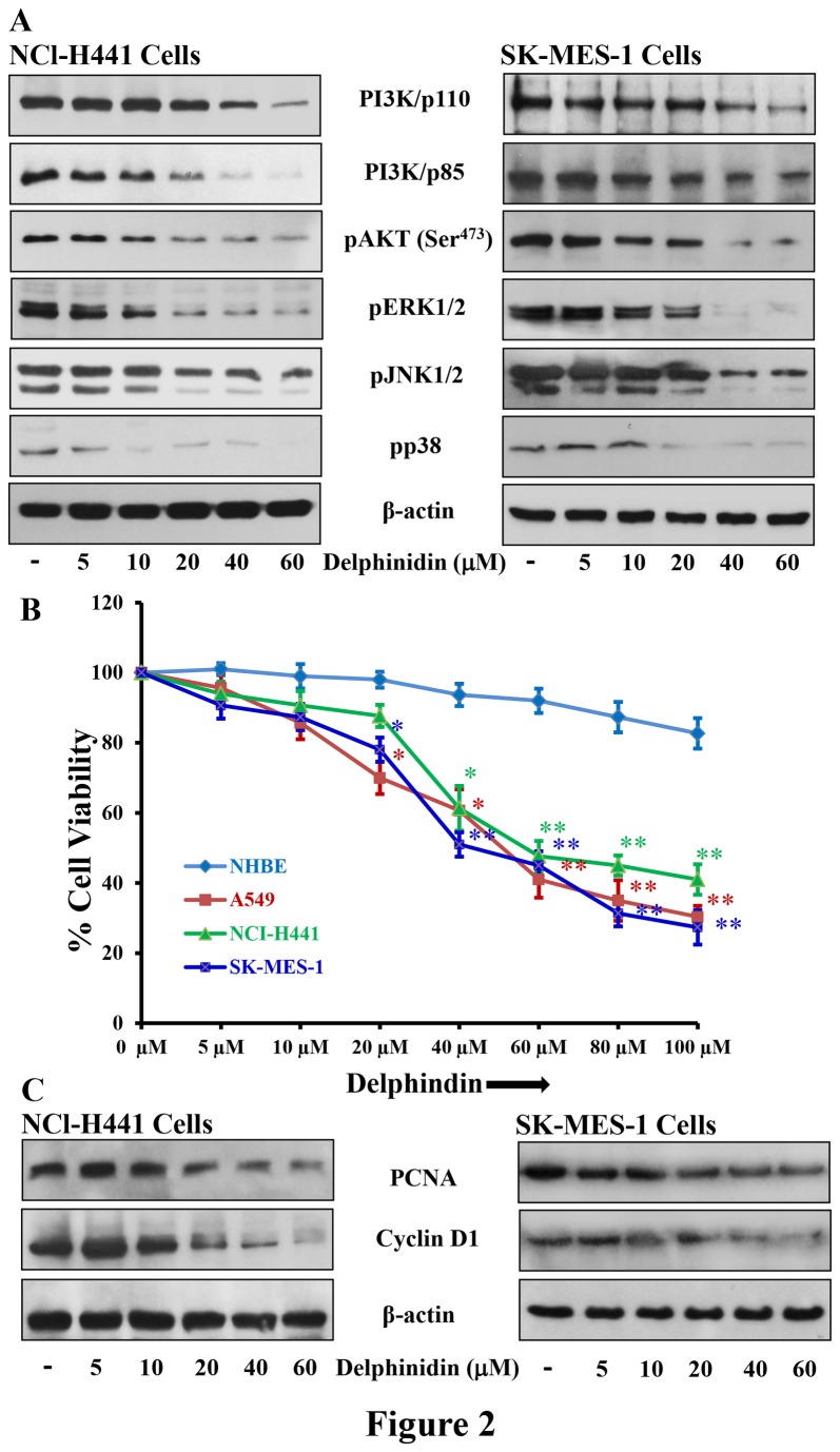
Figure 2. Effect of delphinidin treatment on cell viability protein expression of PI3K, phosphorylation of AKT, MAPKs and expression of PCNA and cyclinD1 in NSCLC cells.
[A] NCI-H441 and SK-MES-1 cells were treated with 5-60 µM delphindin for 48 hrs to determine its effect on protein expression of PI3K, phosphorylation of AKT and MAPKs. After treatment cells were harvested, cell lysates were prepared and protein was subjected to SDS-PAGE followed by immunoblot analysis and chemiluminescence detection. Equal loading of protein was confirmed by stripping the immunoblot and reprobing it for β-actin. The immunoblots shown here are from a representative experiment repeated three times with similar results. [B] Cell viability of A549, NCI-H441, SK-MES-1 and NHBE cells treated with 5-100 µM of delphinidin for 48 hrs, was determined by MTT assay as described in “Materials and Methods”. Data shown are mean ± SEM of three separate experiments in which each treatment was repeated in 10 wells. [C] NCI-H441 and SK-MES-1 cells were treated with 5-60 µM delphindin for 48 hrs to determine its effect on protein expression of PCNA and cyclinD1. After treatment cells were harvested, cell lysates were prepared and protein was subjected to SDS-PAGE followed by immunoblot analysis and chemiluminescence detection. Equal loading of protein was confirmed by stripping the immunoblot and reprobing it for β-actin. The immunoblots shown here are from a representative experiment repeated three times with similar results.

