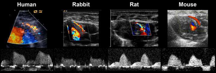
Fig. 3.
Representative ultrasound imaging of the LCA in human, rabbit, rat and mouse. Upper panel: Color Doppler is used for identification of the LCA. In the present imaging windows, the LCA is shown as bright red-yellow streaks. White arrows indicate the site of the LCA where pulsed Doppler is used to measure coronary blood flow velocity. Lower panel: corresponding pulsed wave Doppler signals for measurements of coronary blood flow velocity. Similar flow velocity patterns are evident between the species with minor flow signal during systole and a major flow signal during diastole. Flow velocity on y-axis (maximum velocity 100 cm/s in these images), time on x-axis

