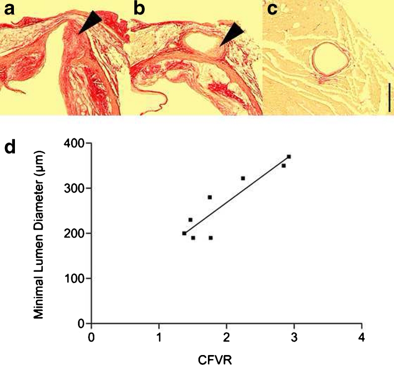
Fig. 5.
Evidence of coronary artery atherosclerosis in the proximal segment (a, b) but not the mid-segment (c) of the left coronary artery in low-density lipoprotein receptor gene-deficient mice. A correlation was evident between minimal lumen diameter and CFVR (d) as imaged with high-resolution ultrasound. Arrowheads indicate coronary lesions. Scale bar = 200 μm. Adapted from [72] with permission

