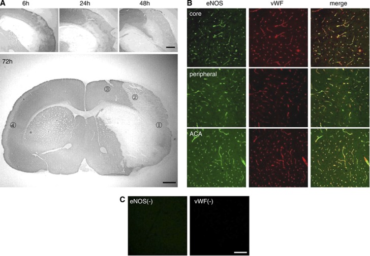
Figure 1.
Ischemic lesions and endothelial nitric oxide synthase (eNOS) expression after transient focal brain ischemia. (A) Microtubule-associated protein immunostaining in the ischemic brain. The ischemic lesion in the brain cortex gradually expanded from the middle cerebral artery (MCA) core to the MCA peripheral area. The bottom panel shows the ischemic lesion 72 hours after ischemia and the regions of interest for assessment; (1) Middle cerebral artery core, (2) MCA peripheral area, (3) anterior cerebral artery (ACA) area, (4) contralateral side. (B) Double immunostaining of eNOS (green) and von Willebrand factor (vWF) (red) in the MCA core, MCA peripheral area, and ACA area. The eNOS signals were mainly localized in the brain microvessels. (C) Immunohistochemistry without primary antibodies showed specificity of eNOS and vWF signals. Scale bar, 1 mm in panel A, 100 μm in panels B and C.

