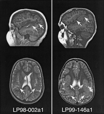
Figure 2.
MRI scans of patients LP98-002a1 and LP99-146a1, showing sagittal T1 (top) and axial T2 (bottom) images for both patients. The images show bilateral extended sylvian fissures lined by polymicrogyric cortex, showing characteristic stippling of the gray-white junction and an irregular, overfolded cortical ribbon (arrows).

