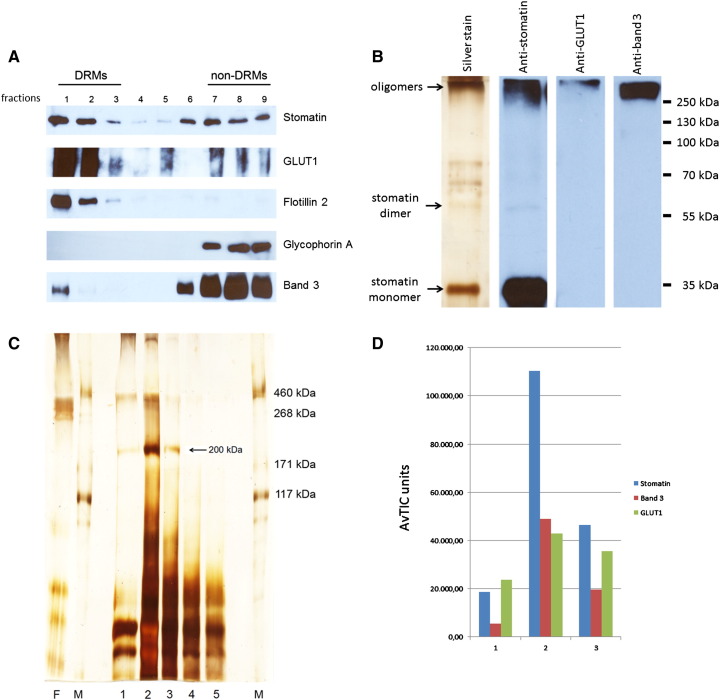
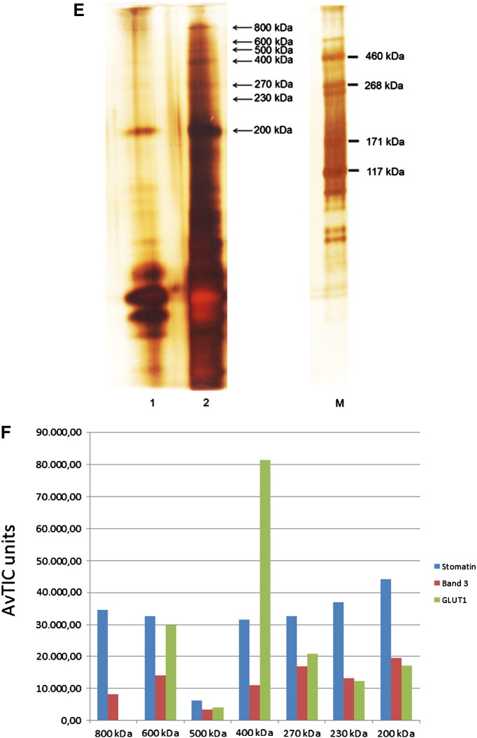
Fig. 5.
Preparation and analysis of EGS-cross-linked high molecular stomatin-complexes in DRMs. (A) Normal ghosts were treated with cold Triton X-100, mixed with alkaline sucrose, and subjected to flotation. Gradient fractions were analysed by Western blotting as indicated. To prevent GLUT1 overstaining in the non-DRM dense fractions 6–9, these fractions were diluted 1:50. Glycophorin A was used as a non-raft marker. (B) Combined DRM fractions 1 + 2 were incubated with 8 μM EGS, quenched, solubilised with SDS, adjusted to 1% Triton X-100, and stomatin-complexes were immunopurified. Elution fraction 1 was analysed by mini-gel 10% SDS-PAGE/silver staining and Western blotting as indicated. High-molecular complexes are visible. Note the major band of non-cross-linked stomatin. (C) Flow-through and elution fractions 1–5 were analysed by 7% SDS-PAGE/silver staining and mass spectrometry of excised 200 kDa bands (fractions 1–3), as indicated. (D) Semi-quantitative MS-analysis shows the composition of the three 200 kDa complexes eluted in fractions 1–3, respectively, reflecting the mass distribution in the fractions. (E) High molecular stomatin-complexes were separated on a 4–12% gradient gel, excised, as indicated, and analysed by MS. (F) Semi-quantitative MS-analysis shows that the region between 200 and 800 kDa contains stomatin-complexes of varying composition. Whereas stomatin is the major constituent of most complexes, the 400 kDa complex contains GLUT1 as major component. Data are compared by AvTIC units. F, flow-through; 1–5, respective elution fractions; M, marker.


