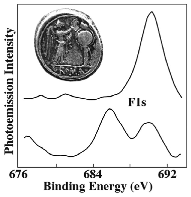
Figure 12.
Use of photoelectron spectromicroscopy to study the microchemical composition of a Roman coin (the optical microscopy image). The local spectra taken after sputtering reveal the unexpected presence of fluorine below the surface. Data extracted from [20].

