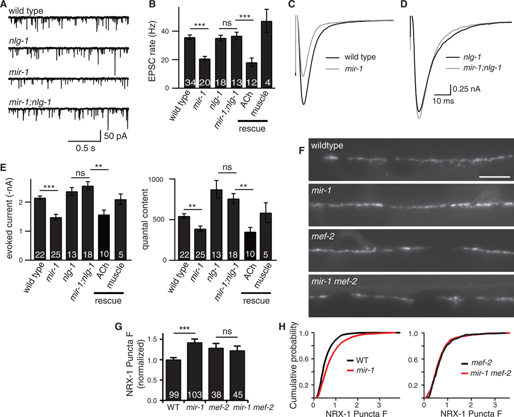
Figure 1. The retrograde signal was induced in mir-1 mutants and inactivated in nlg-1 mutants.
Endogenous (A-B) and evoked EPSCs (C-E) were recorded from adult body wall muscles. Representative traces of endogenous EPSCs (A), averaged evoked responses (C-D), and summary data (B,E) are shown. The effects of miR-1 on EPSC rate and quantal content were eliminated in mir-1; nlg-1 double mutants. (B,E) Retrograde inhibition was re-instated in mir-1; nlg-1 double mutants by nlg-1 transgenes expressed in cholinergic motor neurons (ACh rescue) but not by those expressed in body muscles (muscle rescue). (F-H) Expression of GFP-tagged NRX-1 in body muscles was analyzed. NRX-1 puncta fluorescence in the dorsal cord was significantly increased in mir-1 mutants. This effect was eliminated in mir-1 mef-2 double mutants. Representative images (F), mean (G), and cumulative probability distributions (H) of puncta intensity are shown. Values that differ significantly are indicated (***, p <0.001; **, p <0.01; ns, not significant). The number of animals analyzed is indicated for each genotype. Error bars indicate SEM. Scale bar indicates 5 µm.

