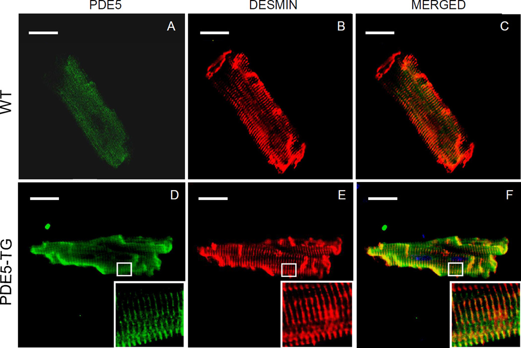
Figure 3. Localization of PDE5 in cardiomyocytes.
Cardiomyocytes isolated from adult WT (A–C) and PDE5-TG (D–F) mice were stained with fluorescently-labeled secondary antibodies against antibodies recognizing PDE5 (green, A and D) and desmin (red, B and E) and were scanned using confocal laser microscopy. The merged view demonstrates that overexpressed PDE5 was predominantly localized to Z-bands (yellow, C and F). Scale bars indicate 20-µm.

