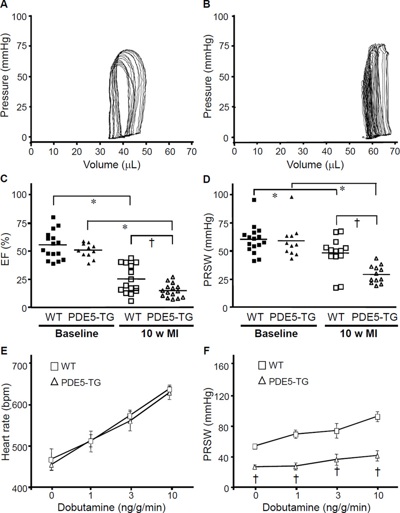
Figure 5. Hemodynamic measurements at baseline and 10 weeks after MI.
Representative pressure-volume loops from WT (A) and PDE5-TG (B) mice 10 weeks after LAD occlusion. Note the rightward shift of the pressure-volume tracings obtained from PDE5-TG. Ejection fraction (C) and preload-recruitable stroke work (D) significantly decreased 10 weeks after MI in both genotypes and to a greater extent in PDE5-TG (*P<0.05 versus baseline; †P<0.05 versus WT). The dobutamine-induced increase in heart rate was similar in both genotypes (E), whereas the increase in PRSW was blunted in PDE5-TG mice (F). †P<0.0001 versus WT.

