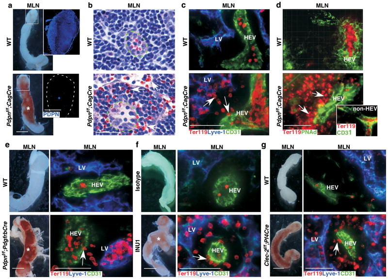
Figure 1. Loss of FRC PDPN or platelet CLEC-2 leads to spontaneous mucosal LN bleeding.
a, Gross morphology of MLNs. Insets contain montages of confocal images of MLN cryosections showing PDPN expression in WT and Pdpnf/f;CagCre mice. b, Images of H&E–stained MLN sections. Arrows indicate extravasated RBCs outside HEVs (dashed line) of Pdpnf/f;CagCre mice. c, Confocal images of MLNs from WT and Pdpnf/f;CagCre mice reveal extravasated RBCs (arrows) outside HEVs, some of which are picked up by lymphatic vessels (LVs). Ter119 indicates RBCs. CD31 marks endothelial cells. Lyve-1 marks LVs. d, Immunostaining of MLN cryosections using HEV-specific marker PNAd. Inset shows that no bleeding occurred around CD31+/PNAd− non-HEV vessels in MLNs. e, Gross morphology and confocal images of MLN cryosections from WT and Pdpnf/f;PdgfrbCre stained for Ter119, Lyve-1, and CD31. f, Gross morphology and confocal images of MLNs from P15 WT mice treated with isotype control rat IgG1κ or the CLEC-2 depleting antibody, INU1. g, Gross morphology and confocal images of MLN cryosections from WT and Clec-2f/f;Pf4Cre mice stained for Ter119, Lyve-1, and CD31. Data are representatives of ≥ 12 mice/group. Scale bars, 2 mm (gross images), 50 μm (b, and confocal images). Asterisk indicates bleeding in the LN. Arrows indicate extravasated RBCs. Tissues were from 1-month-old mice unless otherwise specified.

