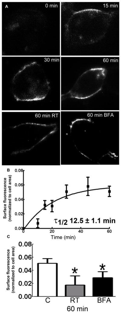
Figure 2.
Insertion of α1β3t-γ2 subunit-containing receptors in HEK293 cells. A: Representative images showing surface α-BT fluorescence in the cells incubated at 37°C for 0, 15, 30 and 60 min, or at RT (panel RT), or with brefeldin-A (5 μg/ml) for 60 min at 37°C (panel BFA). B: The α-BT surface fluorescence was normalized to the cell area, and plotted against the time of incubation at 37°C. C: As a control α-BT fluorescence corresponding to newly inserted receptors was also studied in the cells incubated at RT (gray) or with 5 μg/ml brefeldin-A (black, BFA) for 60 min. The α-BT fluorescence in the cells incubated at 37°C (white) is also plotted for comparison. *P<0.05 vs control (cells incubated at 37°C).

