Figure 1.

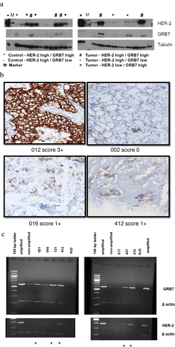
GRB7 and HER-2 expression in frozen breast tumor tissues. a GRB7 and HER-2 Western Blot Analysis. Densitometric analysis is presented in Additional file 1: Table S1. b HER-2 IHC testing of HER-2 FISH positive tumors which do not over-express HER-2 protein by Western blot analysis. Sample 012 is positive control with 3+ HER-2 over-expression; sample 002 is negative control with 0 HER-2 expression. Magnification bars represent 50 micron. c GRB7 and HER2 mRNA expression by multiplex RT-PCR. Top panel: GRB7 and β-actin, Bottom panel: HER-2 and β-actin. Amplified: positive control with HER-2 and GRB 7 over-expression. Non-amplified: negative control - without either HER-2 or GRB7 over-expression. H2O: negative control without RT product. X: 4 tumors over-expressing GRB7 but not HER-2. O: 1 tumor over-expressing both GRB7 and HER-2.
