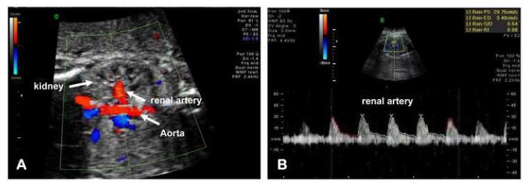
Figure 3. Technique for evaluation and assessment of the fetal renal artery blood flow.
(A) A frontal plane image of the fetal abdomen to allow identification of the abdominal aorta and its bifurcation at the level of the fetal kidneys. Renal artery blood flow was sampled within the lumen of the renal artery away from the aorta and before any emergent branches. The velocity waveform was recorded at fast speed with a low pass filter. (B) Representative Doppler flow tracing of the renal artery in a patient with intra-amniotic inflammation.

