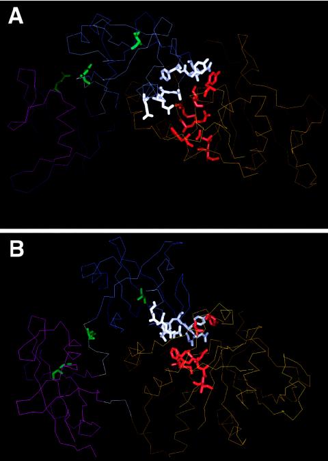
Figure 2.
Location of mutated residues in SHP-2 in the inactive conformation: Cα trace of the N-SH2 (thin blue line), C-SH2 (thin magenta line), and PTP (thin orange line) domains; and N-SH2/C-SH2 and C-SH2/PTP linkers (thin gray lines), according to Hof et al. (1998). The views represented in panels A and B are orthogonal to each other. Mutated residues are indicated with their side chains as thick lines (N-SH2 residues located in or close to the N-SH2/PTP interaction surface are shown in white; linker, C-SH2, and N-SH2-phosphopeptide binding residues are shown in green; and PTP residues located in or close to the N-SH2/PTP interaction surface are shown in red).

