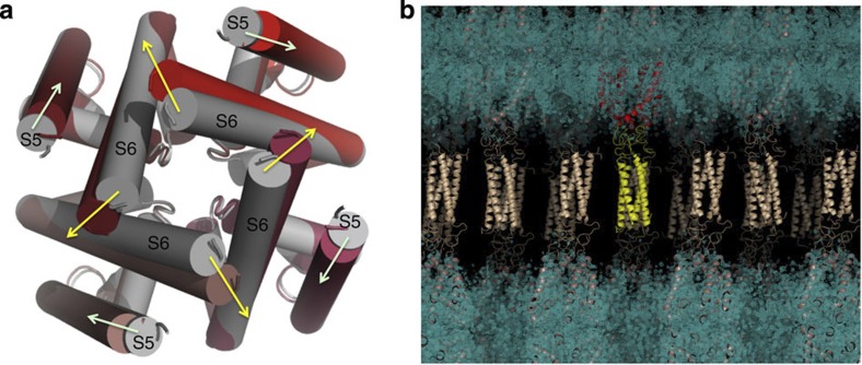
Figure 4. Crystal structure of the NavMs-pore+CTD.
(a) The tetrameric pore structure of NavMs-pore+CTD is in fully open conformation (depicted in cylinder mode, with each monomer in a different shade of red), overlaid for comparison on the closed channel pore structure of the NavAb orthologue (grey). Yellow arrows show the direction of the movement of the end of S6, and light green arrows the movement of the base of S5, between the closed and open pores. (b) Compatibility of the DEER-derived CTD structure with the crystal structure/packing. Electron density map (in blue) overlaid by the structure (in ribbon representation) of the pore domain, with the DEER-defined CTD structure fit into the ‘disordered’ region between ordered tetramers in the crystal lattice. For clarity, one tetramer is depicted with its pore domain in red and its CTD in yellow; the others are in cream.

