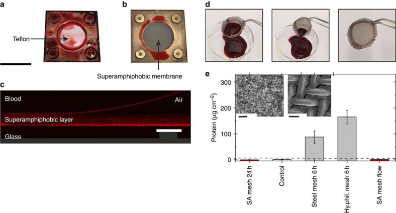
Figure 3. Adsorption of blood on superamphiphobic membranes.
Whole human blood was pumped through a flow cell equipped with two teflon layers (a) or with two superamphiphobic meshes (b). Afterwards the cells were opened and the pictures were taken. (c) Vertical cross-section of a drop of blood on a superamphiphobic layer imaged with a confocal microscope in a reflection mode (supporting information). Reflected light from the superamphiphobic layer, from the blood-air and from the glass-air interface shows that blood does not wet the membrane. (d) Whole human blood rinsed out of a superamphiphobically coated basket made of a steel mesh (Supplementary Movie 1). (e) Results of the protein adsorption assay. The amount of protein adsorbed was measured for (from left to right) the following: a superamphiphobically coated steel mesh after 24 h exposure to non-flowing blood; a control superamphiphobic mesh, which had never been in contact with blood; a bare, uncoated steel mesh after exposing it to non-flowing blood for 6 h; a coated but unfluorinated steel mesh after 6 h exposure to non-flowing blood (Hy.phil. mesh). In addition, a superamphiphobic steel mesh was exposed to a blood flow (5.12 ml min−1) for 3 h. Washing of the samples (three times with 3 ml PBS) removed blood that did not adhere to the surface. Error bars are based upon the root mean square deviation. The horizontal dashed line is the detection limit of 6 μg cm−2. Inset: (left) SEM image of a superamphiphobic membrane after 48 h exposure to whole human blood; (right) SEM image of the corresponding metal mesh after 2 h exposure to whole human blood. Scale bars, 2 mm (a,b), 50 μm (c), 10 μm (e, left) and 50 μm (e, right).

