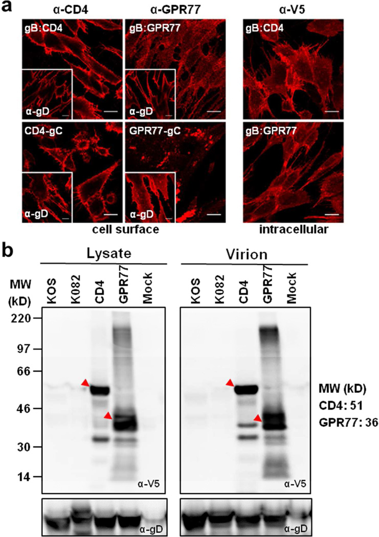
Figure 2.
Expression of CD4 and GPR77. a. Confocal analysis of HFT cells infected with the recombinant viruses demonstrating cell surface expression of CD4 and GPR77. The cell surface expression of gD is shown for similar infected cells in the insets of each panel. Intracellular distribution of CD4 and GPR77 expressed from the gB promoter was visualized by staining with anti-V5 antibodies following permeabilization of cells. Scale bars: 200 microns. b. Expression of CD4 and GPR77 in infected cell lysates and incorporation of these proteins in mature virions was confirmed by western blot analysis using anti-V5 antibody.

