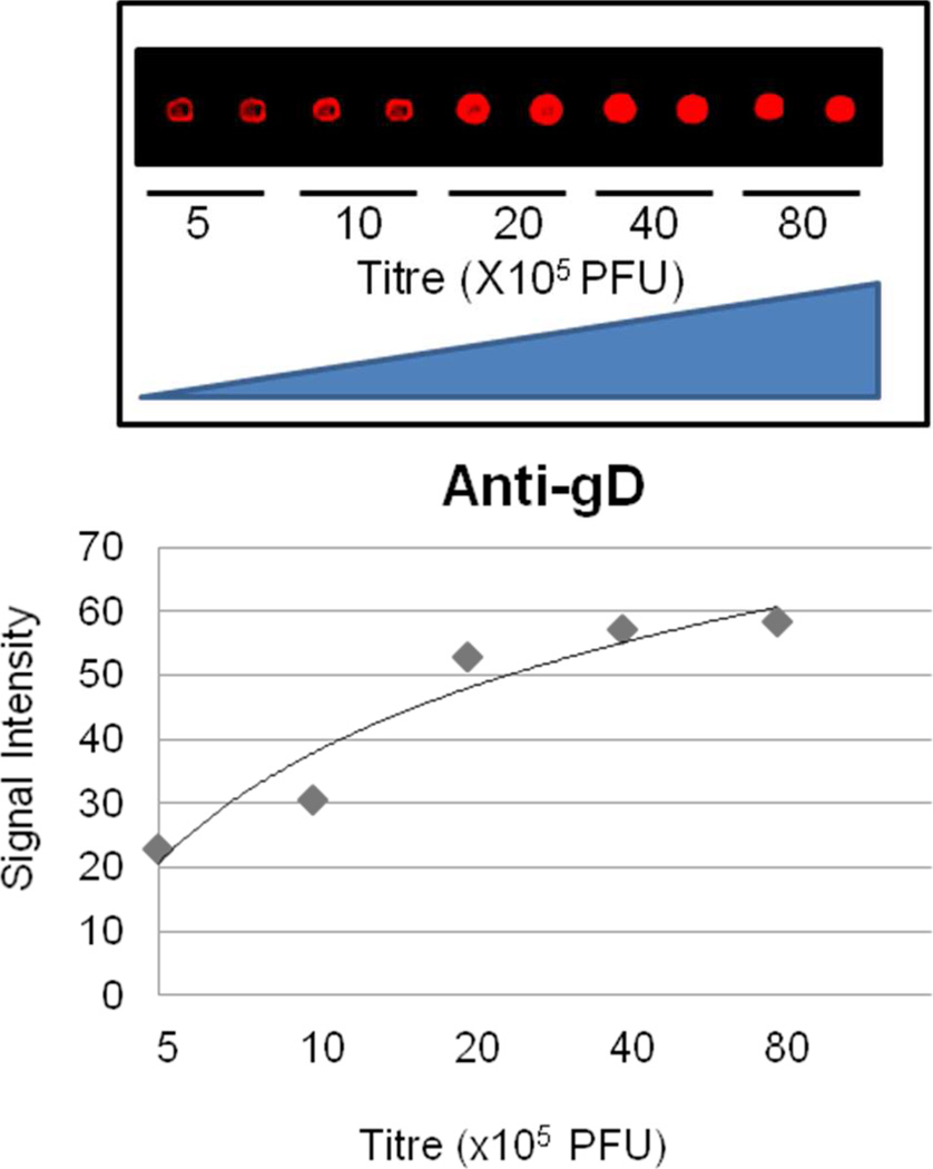
Figure 4.
Wild-type HSV-1 virions were printed on the FAST glass surface at different concentrations. Each spot represents 2.1 nl of virus suspension. The virions were detected using an anti-gD antibody against the ectodomain of this molecule. 500,000 virions (KOS plaque forming units) per spot can be easily detected by the anti-gD antibody on the VirD Arrays. The fluorescence signal begins to reach saturation after the number of virus particles printed increases to >4,000,000 virions.

